Slate magazine had a slideshow by Daniel Engber a little more than a week ago on unusual laboratory animals and why they're important. The slideshow was prompted by Engber's observation that mice and rats make up an enormous proportion of all lab animals, perhaps limiting what we can conclude from experimental results and narrowing our perspective on what questions to ask. In short, scientists need to start thinking outside the box when it comes to model organisms. Engber lists fourteen animals, some of which have already given important clues to specific questions. I will mention some of those here.
1. The squid: the squid peaked in importance in the 1950's, when Hodgkin and Huxley got the idea to use its giant axon (up to 1mm diameter) to study properties of the action potential. The large diameter made it possible for them to insert micro-electrodes directly into the intracellular space of the axon, thereby measuring the flow of ions across the membrane during various stages of the action potential or under different extracellular ionic concentrations. This work resulted in Hodgkin and Huxley's mathematical model of AP generation, which earned them the 1963 Nobel Prize. 
The squid giant axon (not "giant squid axon") was used primarily because its large diameter made it possible for researchers to stick electrodes into the lumen. These days, better equipment and techniques allow scientists to record electrical activity of neurons with intracellular electrodes placed into cell bodies with widths in the range tens of microns in slice preparations or even awake behaving animals!
2. Xenopus laevis frogs are used for the eggs females produce, oocytes, which can be around 1mm in diameter. As with the squid giant axon, these cells were first used because their large size makes them easy to handle. 
Xenopus oocytes contain machinery for protein production, which scientists can hijack by injecting DNA or RNA coding a desired protein. Once injected with DNA or RNA, the oocyte starts making protein. Oocytes are typically used in electrophysiological experiments on the function of particular neurotransmitter- or voltage-gated ion channels, like the GABA-A receptor.
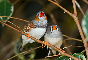
3. The zebra finch and other songbirds are great models for motor learning/planning and language acquisition. Adult male Zebra finches sing the same song - a highly structured, complex sequence of sounds that requires equally sophisticated motor control - throughout their lives (hundreds of times per day); how the brain codes for such learned sequences and maintains them for years is a question of great interest, and arguably more amenable to scientific study than motor control in primates (the typical model for such questions), whose movement repertoire is much more diverse and variable. 
Other birds don't sing the same tune every time, but instead combine syllables or motifs into songs that differ in composition each time. Canaries have a rich repertoire that may provide clues into sequence and even syntax learning. Their songs contain non-random sequences of syllables, each of which determines what syllable comes next. So any given syllable transition point in a Canary song depends on what syllable came before. Even cooler, Bengalese finches were recently shown to be able to spontaneously discriminate among songs of variable syntactic structure, indicating that birds have not only the ability to produce songs with hierarchical syllable structure, but to perceive such structure too.
Other model organisms Engber mentions are the zebrafish, traditionally used in genetics experiments, has recently been added to the list of species with an optogenetics toolkit (others include flies, mice and primates); the sea slug Aplysia - used famously by Eric Kandel to demonstrate the concept of long term potentiation, LTP, and the molecular principles behind it; the prairie vole - first demonstration of molecular principles behind "monogamous" relationships; the fruit fly, used for everything genetic because its generations are so short, actually accounts for a small fraction of all papers published since the 1950's, according to Engber's analysis.
We do need out of the box thinking in asking the right questions in science. Choosing appropriate (or simply different) model organisms could be one way to start. The animals listed above are good examples of the results that were achieved by doing so. Archaebacteria also come to mind, since they were the first organisms to contribute to the booming field of optogenetics. But using the same organism to study a given scientific problem is still a necessary way to standardize all the research coming out every day. Tons of data would be uninterpretable if every team used a different organism to perform different experiments. What will the next animal of choice be for neuroscience? Something with decent intelligence, complex neurophysiology, yet not so complex that it becomes criminal to keep it in a cage. Perhaps corvids, with their amazing cognition despite the lack of a six-layer neocortex?


